What the Cell
B2.1

Membranes and membrane transport

HL extension
3 hours7 statementsTextbook pp. 237–243

Guiding questions

  1. How do lipid and protein molecules come together to form membranes?
  2. What decides whether a substance can cross a membrane?

Slides

Slide 1 of 17

Notes

Written for this site from the class slides, with textbook page references for each statement. The chapter is on pages 237–243 of the course textbook. Image credits are on slide 17.

Membrane fluidity

B2.1.11

Fatty acids and fluidity

p. 237

Saturated fatty acid tails are straight and pack closely, so they make a membrane less fluid. Unsaturated tails are kinked at their double bonds, cannot pack closely and keep the membrane fluid at lower temperatures. Membranes must be fluid enough for proteins to move and work, but not so fluid that they leak. Organisms such as the lake sturgeon raise the share of unsaturated fatty acids when the water gets colder: homeoviscous adaptation.
B2.1.12

Cholesterol

pp. 237–238

In animal membranes, cholesterol sits with its polar -OH group next to the phosphate heads and its non-polar rings and tail among the fatty acid tails. It acts as a fluidity buffer: at low temperature it keeps phospholipids apart so the membrane stays fluid; at high temperature it restricts their movement so the membrane is less fluid and less leaky.

B2.1.13

Vesicles form and fuse

pp. 238–239

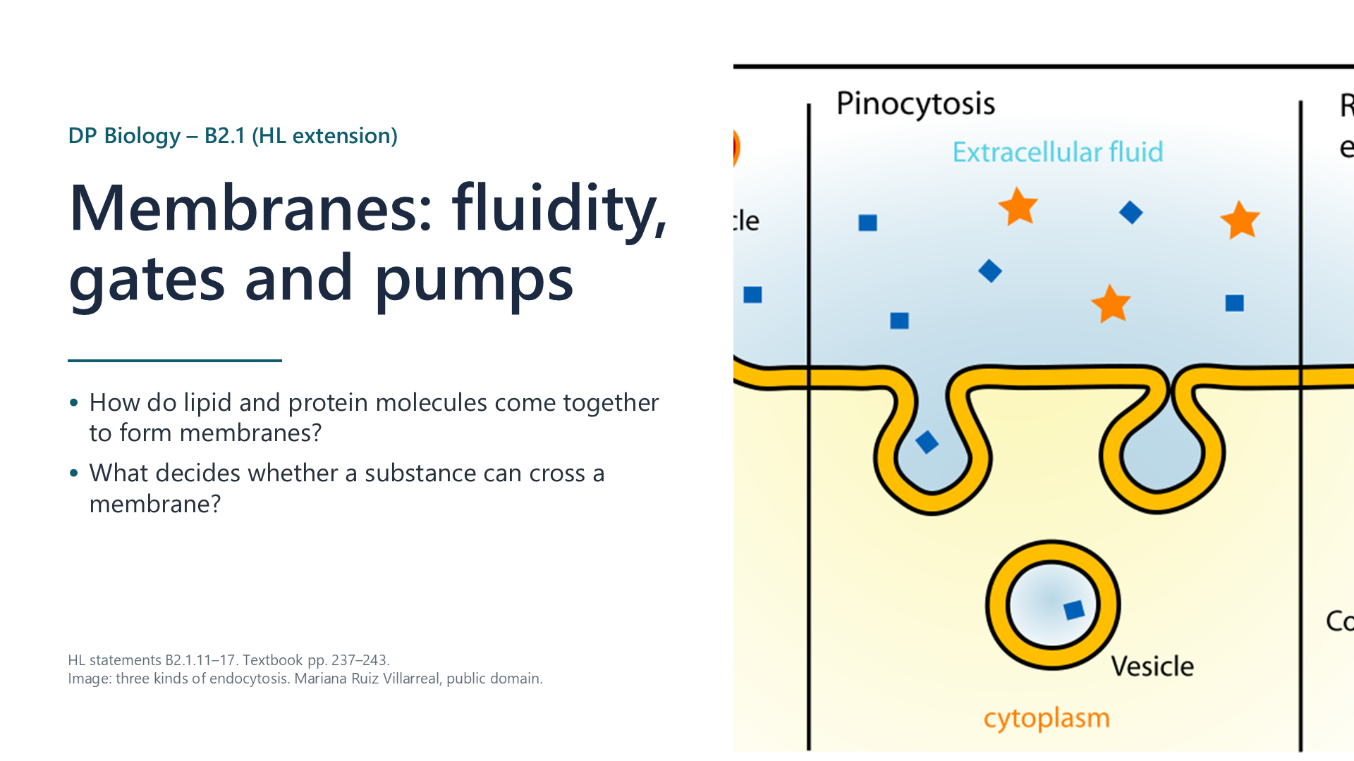
Because phospholipids can flow, a membrane can pinch inwards to make a vesicle (endocytosis: phagocytosis of solids, pinocytosis of liquids) and a vesicle can fuse with the plasma membrane (exocytosis) without leaving a hole. Both need ATP. Macrophages engulf old red blood cells; plasma cells secrete antibodies and neurons release neurotransmitters by exocytosis.

Channels, pumps and cotransporters

B2.1.14

Gated ion channels

pp. 239–241

Gated channels open and close, letting ions diffuse down their gradients. Voltage-gated Na+ and K+ channels in axons open when the membrane potential changes; a K+ channel is then plugged by a “ball and chain” until the membrane repolarizes. Neurotransmitter-gated channels, such as the nicotinic acetylcholine receptor, open when acetylcholine binds, letting Na+ into the post-synaptic cell.

B2.1.15

The sodium-potassium pump

pp. 241–242
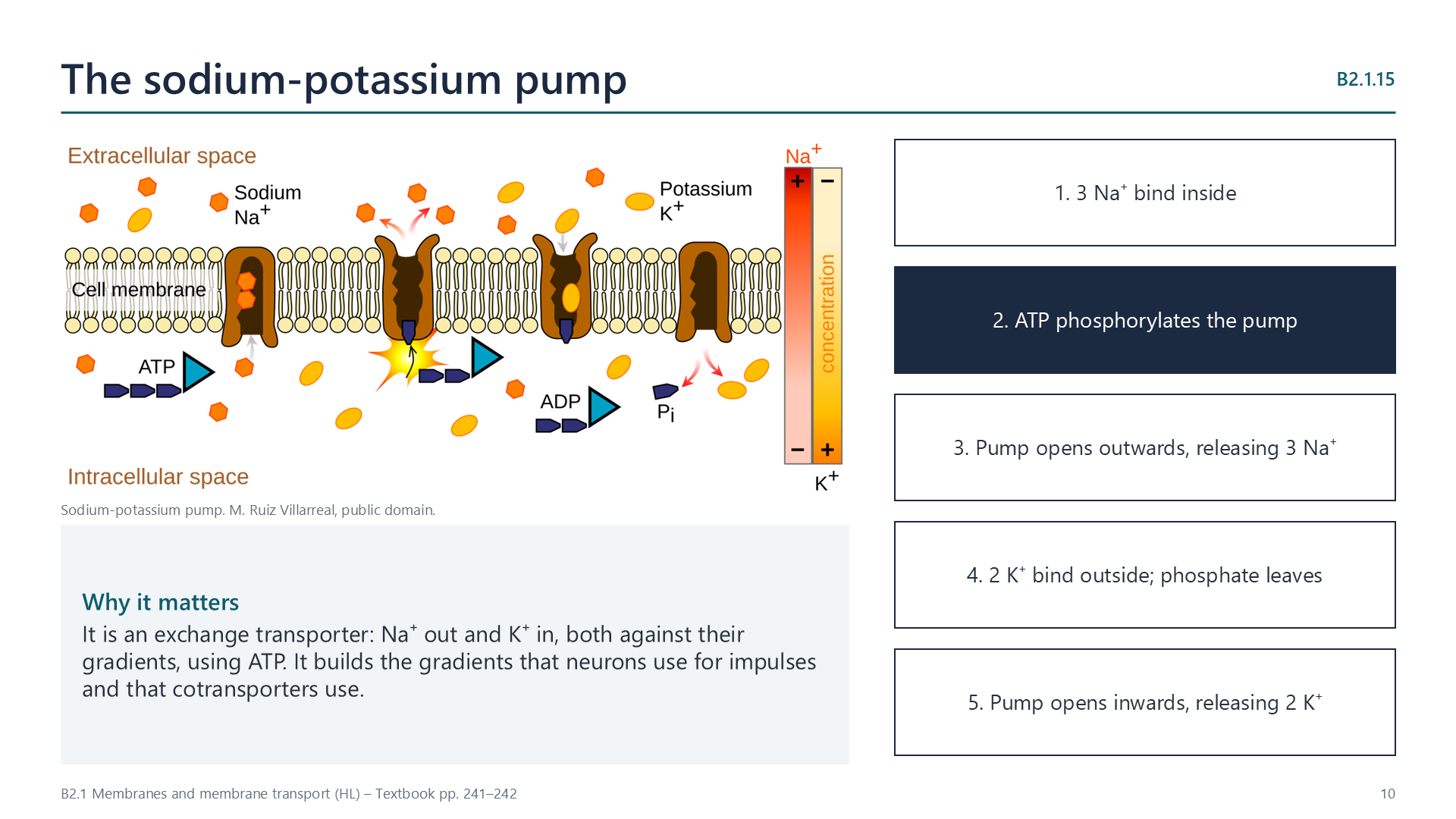
Slide: diagram of the sodium-potassium pump beside its five steps

An exchange transporter. Three Na+ bind inside; ATP phosphorylates the pump, which opens outwards and releases them; two K+ bind outside; the phosphate leaves and the pump opens inwards, releasing K+. Both ions move against their gradients, building the gradients used by neurons and cotransporters.

B2.1.16

Indirect active transport of glucose

p. 242

In the small intestine and kidney tubules, Na+/K+ pumps keep Na+ low inside the cell. Na+ diffuses back in through a sodium-dependent glucose cotransporter, carrying glucose with it (two Na+ per glucose), even against the glucose gradient. The cotransporter uses no ATP itself, so this is indirect active transport. Glucose then leaves the cell by facilitated diffusion into the blood.

Tissues

B2.1.17

Cell adhesion

p. 243

Cell-adhesion molecules (CAMs) are membrane proteins that bind cells to each other (usually cadherins) or to the extracellular matrix (usually integrins). They form junctions: tight junctions seal epithelia, anchoring junctions give strength and gap junctions link the cytoplasm of neighbouring cells. Plant cells rely instead on interlocking walls and plasmodesmata.

Videos

Select a video to play it here. Select it again to close it.

Note-taking sheet

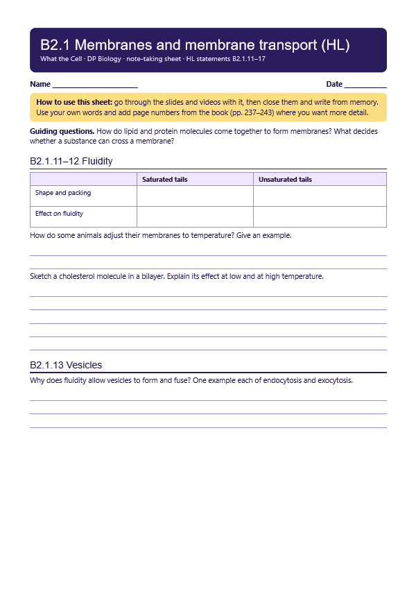
Print this guide and complete it as you work through the slides and videos. It asks for your own explanations, so close the notes while you try each section.

First page of the note-taking sheet

Quick self-check

The self-check needs JavaScript to run.

Practice questions

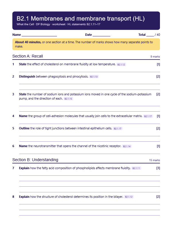
A printable worksheet covering all seven statements, from quick recall to data questions and a longer answer. It has 40 marks and takes about 40 minutes, or you can do one section at a time.

Your teacher has the mark scheme.

First page of the worksheet

Practical

Temperature and membrane permeability

Lab50 minB2.1.11, B2.1.12

You will need: Beetroot cylinders, water baths from 20 to 70 C, test tubes, colorimeter or colour chart

  1. Rinse the beetroot until the water runs clear
  2. Leave equal pieces in water at each temperature for 20 minutes
  3. Compare the colour of the water
  4. Plot the results, and explain them using membrane fluidity

Safety: Take care with hot water and with cutting tools. Beetroot stains.

Carry this out with your teacher's supervision and follow your school's lab rules.

Nature of science and TOK

Syllabus statements

Summarised in our own words. Use each code to find the exact wording in your textbook or the IB subject guide.

  • B2.1.11How the fatty acids in a bilayer affect its fluidityp. 237
  • B2.1.12How cholesterol affects membrane fluidity in animal cellsp. 237
  • B2.1.13How fluid membranes allow vesicles to form and fusep. 238
  • B2.1.14Neuron ion channels that open and close (gated channels)p. 239
  • B2.1.15The sodium-potassium pump as an exchange transporterp. 241
  • B2.1.16Indirect active transport: how sodium-driven glucose cotransporters workp. 242
  • B2.1.17How cells adhere to one another to build tissuesp. 243

More of B2.1 in this course

© 2026 Akash Parthi Bendran. Please ask before using or sharing any resources from this site: see permission to use.