What the Cell
B2.2

Organelles and compartmentalization

HL extension
2 hours6 statementsTextbook pp. 248–255

Guiding questions

  1. How does the structure of each organelle suit its function?
  2. Why is it useful for cells to have separate compartments?

Slides

Slide 1 of 13

Notes

Written for this site from the class slides, with textbook page references for each statement. The chapter is on pages 248–255 of the course textbook. Image credits are on slide 13.

Organelles that make ATP

B2.2.4

The mitochondrion

pp. 249–250
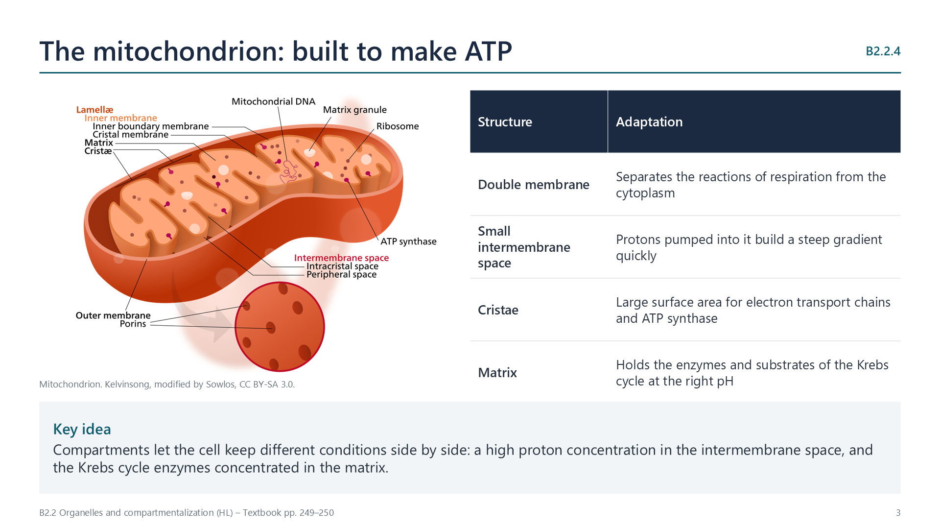
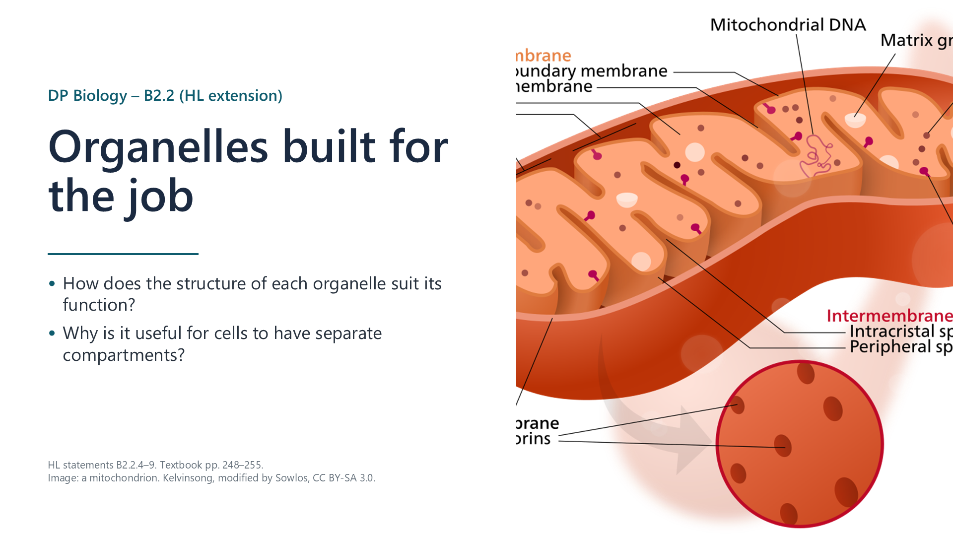
Slide: labelled mitochondrion beside a table of its adaptations

A double membrane separates aerobic respiration from the cytoplasm. The inner membrane is folded into cristae, giving a large surface area for electron transport chains and ATP synthase. Protons are pumped into the small intermembrane space, so a steep gradient builds quickly. The matrix holds the Krebs cycle enzymes and substrates together at a suitable pH.
B2.2.5

The chloroplast

pp. 250–252

Stacked thylakoid membranes (grana) give a large surface area for photosystems, so much light is absorbed. Protons pumped into the small volume inside the thylakoids quickly make a steep gradient for ATP synthase. The stroma holds the Calvin cycle enzymes, such as rubisco, and their substrates, separate from the light-dependent reactions.

B2.2.6

The double nuclear membrane

pp. 252–253

The two membranes of the nuclear envelope keep DNA apart from the cytoplasm. They join at the nuclear pores, which control traffic: mRNA leaves; nucleotides, proteins and ATP enter. The outer membrane is continuous with the rough ER. At the start of mitosis and meiosis the envelope breaks into vesicles, so spindle microtubules can reach the chromosomes; afterwards the vesicles re-form a nucleus around each set.

Making and moving proteins

B2.2.7

Free ribosomes and rough ER

pp. 253–254

Both are 80S ribosomes. Free ribosomes make proteins that stay in the cytoplasm, or are imported into the nucleus, mitochondria or chloroplasts. Ribosomes bound to the rough ER make proteins for secretion, for lysosomes or for the plasma membrane; these enter the ER lumen and travel on in vesicles. Several ribosomes on one mRNA (a polysome) make many copies at once.

B2.2.8

The Golgi apparatus

pp. 254–255

A stack of flattened sacs (cisternae). Vesicles from the rough ER fuse with the side facing the ER. Proteins pass from cisterna to cisterna, where enzymes modify them, for example by adding sugars (glycosylation) or phosphate. These changes label proteins for their destination. Vesicles bud from the far side and carry the proteins to the plasma membrane for secretion, or to lysosomes.

B2.2.9

Vesicles and clathrin

p. 255
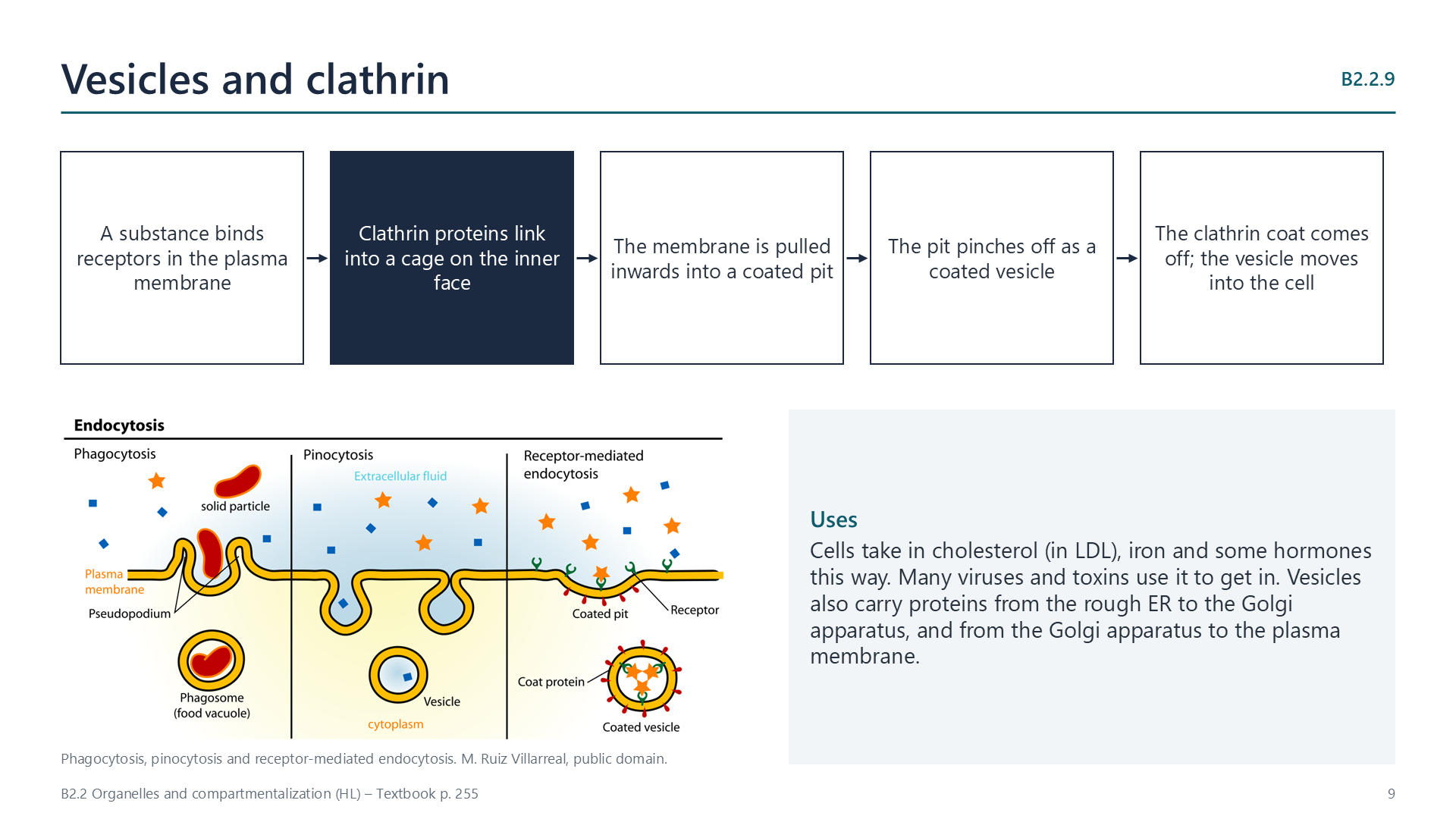
Slide: five steps of clathrin-coated vesicle formation, with a diagram of three kinds of endocytosis

Vesicles are small sacs of membrane that carry materials between compartments and across the plasma membrane. In receptor-mediated endocytosis, a substance binds receptors and clathrin proteins link into a cage on the inner face of the membrane, pulling it into a coated pit that pinches off as a vesicle. Cells take in LDL cholesterol this way, and many viruses use it to enter.

Videos

Select a video to play it here. Select it again to close it.

Note-taking sheet

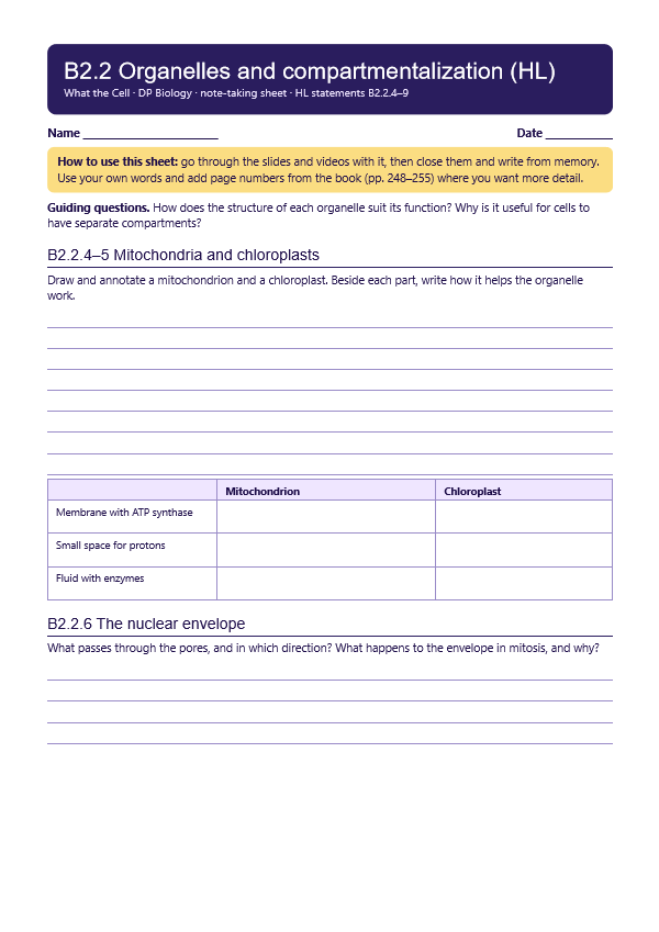
Print this guide and complete it as you work through the slides and videos. It asks for your own explanations, so close the notes while you try each section.

First page of the note-taking sheet

Quick self-check

The self-check needs JavaScript to run.

Practice questions

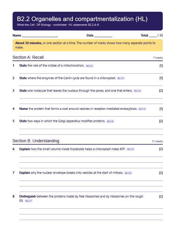
A printable worksheet covering all six statements, from quick recall to data questions and a longer answer. It has 30 marks and takes about 30 minutes, or you can do one section at a time.

Your teacher has the mark scheme.

First page of the worksheet

Practical

Organelles in electron micrographs

Data35 minB2.2.4, B2.2.5

You will need: Public-domain electron micrographs of a mitochondrion and a chloroplast, ruler

  1. Identify and label the membranes and internal structures
  2. Use the scale bar to calculate the actual length
  3. Annotate how each feature suits the organelle's function
  4. Compare the two organelles

Safety: No practical hazards.

Carry this out with your teacher's supervision and follow your school's lab rules.

Nature of science and TOK

Syllabus statements

Summarised in our own words. Use each code to find the exact wording in your textbook or the IB subject guide.

  • B2.2.4How the mitochondrion is adapted for making ATP in aerobic respirationp. 248
  • B2.2.5How the chloroplast is adapted for photosynthesisp. 249
  • B2.2.6What the nucleus gains from having a double membranep. 251
  • B2.2.7The structure and roles of free ribosomes and rough endoplasmic reticulump. 253
  • B2.2.8The structure and role of the Golgi apparatusp. 254
  • B2.2.9The structure and role of vesiclesp. 255

More of B2.2 in this course

© 2026 Akash Parthi Bendran. Please ask before using or sharing any resources from this site: see permission to use.