What the Cell
C2.2

Neural signalling

4 hours7 statementsTextbook pp. 467–478

Guiding questions

  1. How do neurons produce electrical signals and pass them along?
  2. How do neurons affect other cells?

Slides

Slide 1 of 22

Notes

Written for this site from the course textbook and class slides. Use the page numbers to read the book's fuller account. Image credits are on slide 22.

Neurons at rest

C2.2.1

What neurons are

pp. 467–469

The nervous system is made of cells called neurons. The brain and spinal cord form the central nervous system, and peripheral nerves link it with the rest of the body.

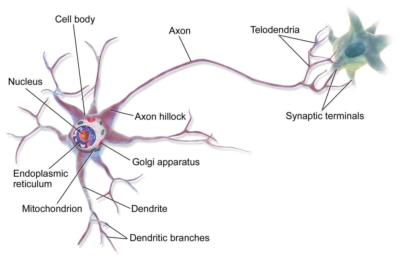
A neuron has a cell body, containing cytoplasm and a nucleus, with long nerve fibres projecting from it. Dendrites are many short fibres that bring impulses to the cell body. An axon is one long fibre that carries impulses away. Electrical impulses travel along these fibres.

Sensory neurons bring signals from receptors, relay neurons connect neurons in the CNS, and motor neurons take signals out to effectors, which are muscles and glands.
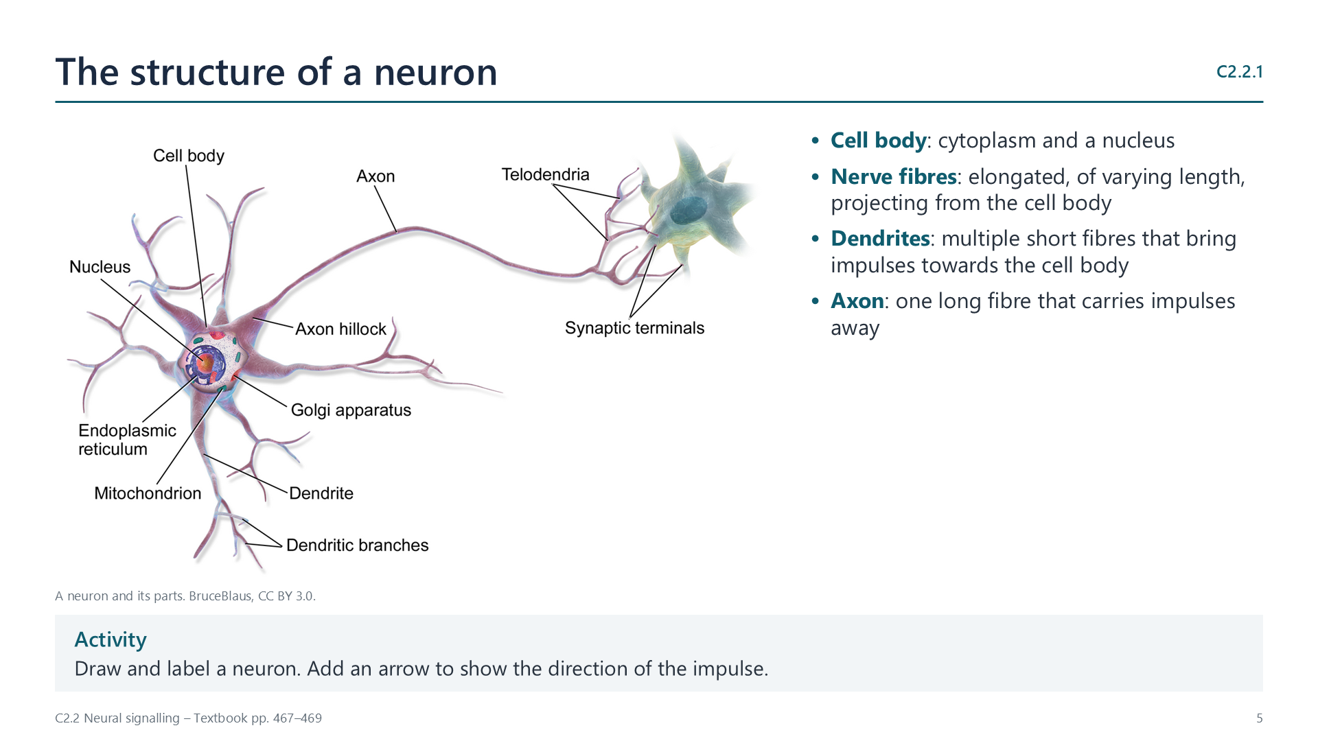
Slide: diagram of a neuron with key parts listed beside it

Watch out: receptors detect stimuli and effectors respond to them.
C2.2.2

How pumping ions sets up the resting potential

pp. 469–470

A membrane potential is a difference in charge across a membrane. A membrane with positive charge on one side and negative on the other is polarized. In a neuron that is not transmitting an impulse, the inside is negative at about −70 mV. This is the resting potential.

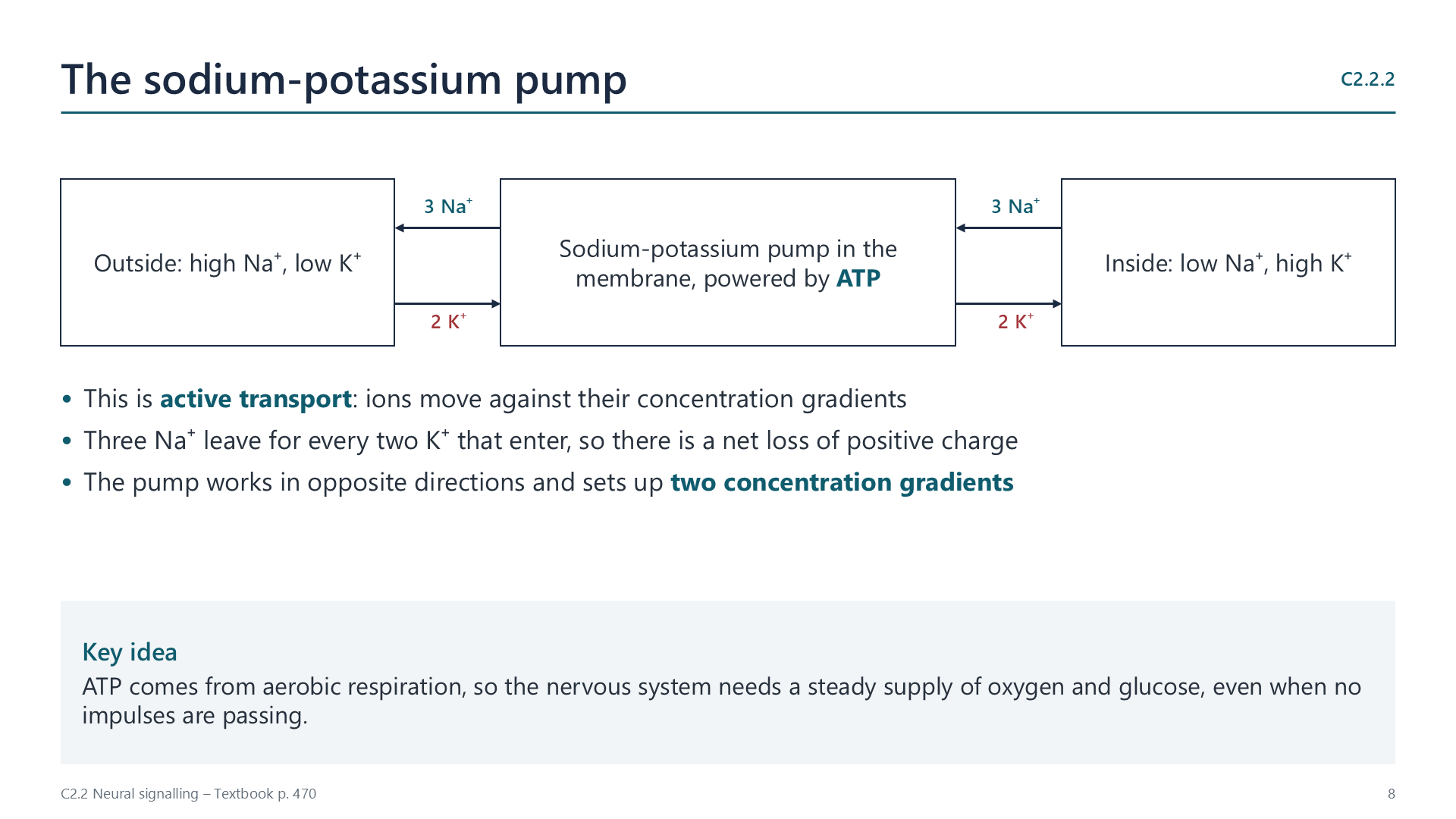
  • The pump: sodium-potassium pumps use ATP to move three Na+ out for every two K+ in. This active transport builds a concentration gradient for each ion, and removes more positive charge than it brings in.
  • Leakage: the membrane is more permeable to K+, so K+ leaks out faster than Na+ leaks in.
  • Proteins: large, negatively charged proteins stay inside the axon.

So a "resting" neuron is not idle. It uses ATP all the time to keep the gradients in place.

Slide: the sodium-potassium pump moving three sodium ions out and two potassium ions in

The impulse

C2.2.3

Nerve impulses: action potentials travelling along nerve fibres

p. 471
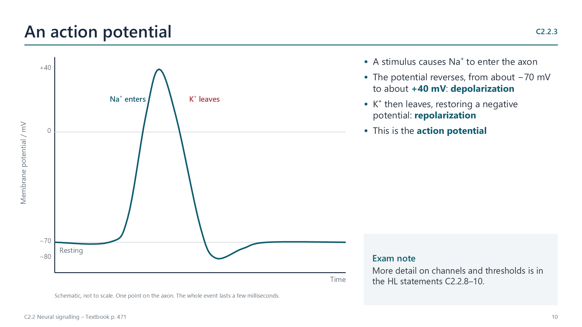
Slide: graph of membrane potential rising from minus 70 to plus 40 millivolts and returning

A stimulus lets positively charged sodium ions flow into the axon, and the potential reverses from about −70 mV to about +40 mV. Potassium ions then flow out, restoring a negative potential. This brief change is an action potential.

An action potential at one point triggers the next region of the fibre, so the impulse is propagated along it. At any one point it lasts only about two milliseconds.

The impulse is electrical because it involves the movement of charged ions, which changes the voltage across the membrane. It is not a flow of electrons along a wire.

C2.2.4

Why nerve impulses travel at different speeds

pp. 472–475
FibreDiameterMyelinTypical speed
Small fibre in a mammal1 µmNoAbout 1 m s−1
Squid giant axonUp to 500 µmNoAbout 25 m s−1
Mammal motor fibreAbout 20 µmYesAbout 100 m s−1

Diameter: a narrow fibre has greater resistance to the flow of ions inside it, so a wider fibre conducts faster. Giant axons help a squid to escape quickly.

Myelin: Schwann cells wrap around the axon, forming a lipid-rich sheath with high electrical resistance. Ions cross the membrane only at the gaps, or nodes of Ranvier, so the impulse jumps from node to node. This is far faster, even in much narrower fibres.

Passing the signal on

C2.2.5

Synapses: junctions with neurons and effectors

p. 476

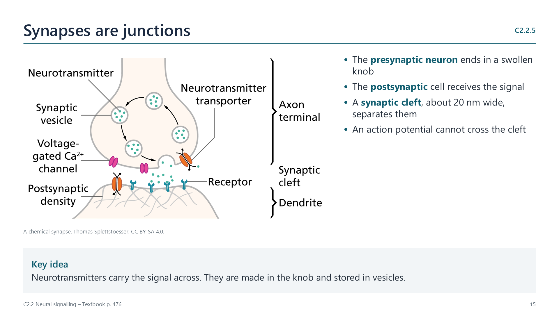
A synapse is a junction between a neuron and another neuron, or between a neuron and an effector, such as a muscle fibre or a gland. The two cells do not touch. A gap, the synaptic cleft, is about 20 nm wide, and an action potential cannot cross it.

Chemical neurotransmitters carry the signal across instead. A signal can cross a typical synapse in only one direction, because vesicles occur only on the presynaptic side and receptors only on the postsynaptic side.

Slide: illustration of a chemical synapse beside key points
C2.2.6

How the presynaptic membrane releases neurotransmitter

p. 477
  1. An action potential arrives and depolarizes the presynaptic membrane.
  2. Calcium channels open, and Ca2+ diffuses into the knob.
  3. Calcium acts as a signalling chemical inside the neuron, making vesicles move to the membrane.
  4. Vesicles fuse with the membrane, releasing neurotransmitter into the cleft.
C2.2.7

How an excitatory postsynaptic potential is produced

pp. 477–478

Use acetylcholine (ACh) as your example. It is used at many synapses, including the neuromuscular junction.

  1. ACh diffuses across the cleft, from high to low concentration.
  2. It binds to specific transmembrane receptors on the postsynaptic membrane.
  3. These receptors are also ion channels. They open, and Na+ flows in.
  4. This depolarizes the membrane: an excitatory postsynaptic potential. If it is large enough, it starts an action potential in the next cell.
  5. The enzyme acetylcholinesterase breaks ACh down to choline and acetate. The channel closes, and the choline is reabsorbed and reused.

Watch out: always say that the neurotransmitter crosses the cleft by diffusion. Remember to mention its removal by an enzyme.

Videos

Select a video to play it here. Select it again to close it.

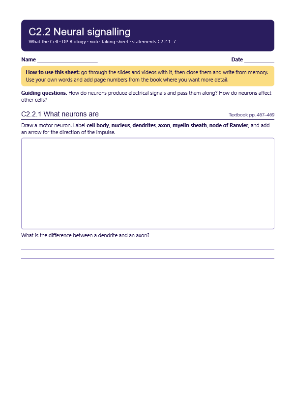
Note-taking sheet

Print this guide and complete it as you work through the slides and videos. It asks for your own explanations, so close the notes while you try each section.

First page of the note-taking sheet

Quick self-check

The self-check needs JavaScript to run.

Practice questions

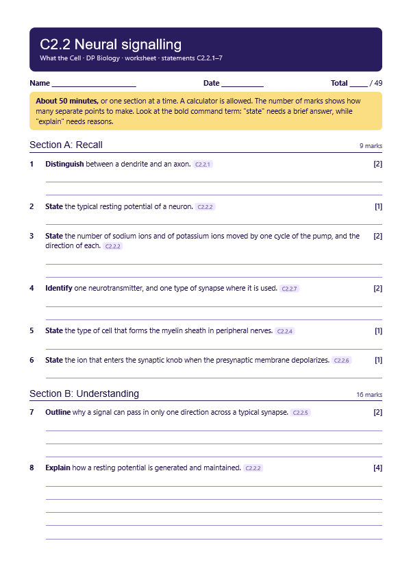
A printable worksheet covering all seven statements, from quick recall to data questions and a longer answer. It has 49 marks and takes about 50 minutes, or you can do one section at a time.

Your teacher has the mark scheme. Past-paper practice: IB Biology SL, November 2021, Paper 1, questions 3 and 28. Ask your teacher for a copy.

First page of the worksheet

Data-based questions

Questions built on realistic experimental data, in the style of Paper 1B and Paper 2. Work them on paper and show your working. 8 marks in total. Your teacher has the mark scheme.

Smelling danger

8 marks
A neuron
Photo: BruceBlaus, CC BY 3.0, Wikimedia Commons.

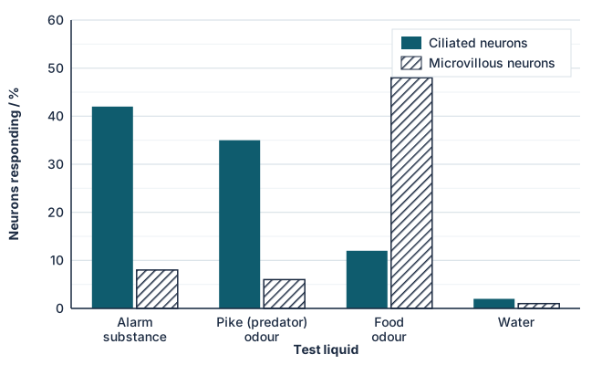
When a fish is injured, its skin releases an alarm substance that warns other fish. Scientists recorded from two types of smell (olfactory) neurons in the noses of zebrafish while washing over one of four test liquids. The graph shows the percentage of neurons of each type that responded with action potentials.

Bar chart of neurons responding. Ciliated neurons: alarm substance 42%, pike odour 35%, food odour 12%, water 2%. Microvillous neurons: 8%, 6%, 48%, 1%.
  1. State the percentage of ciliated neurons that responded to pike odour. [1]
  2. Identify, with a reason, which type of neuron is mainly used to detect danger. [1]
  3. Explain why water was included as one of the test liquids. [1]
  4. Compare the responses of the two types of neuron to alarm substance and food odour. [2]
  5. Explain how the nervous system could tell a strong smell from a weak one, given that action potentials are all-or-nothing. [2]
  6. Suggest an advantage of detecting alarm substance and predator odour with the same neurons. [1]

Practical

Reaction time with a falling ruler

Lab25 minC2.2.1, C2.2.5

You will need: 30 cm ruler, reaction time conversion formula

  1. Partner holds the ruler above your open hand, then drops it without warning
  2. Record the distance fallen before you catch it, for ten trials
  3. Convert distance to time using t = square root of (2d / 9.8), with d in metres
  4. Discuss why the response takes time, using neurons and synapses in your answer

Safety: Low risk. Keep the ruler low and away from faces.

Carry this out with your teacher's supervision and follow your school's lab rules.

Nature of science and TOK

Syllabus statements

Summarised in our own words. Use each code to find the exact wording in your textbook or the IB subject guide.

  • C2.2.1What neurons are: nervous system cells that conduct electrical signalsp. 467
  • C2.2.2How pumping sodium and potassium ions sets up and maintains the resting potentialp. 469
  • C2.2.3Nerve impulses: action potentials travelling along nerve fibresp. 471
  • C2.2.4Why nerve impulses travel at different speedsp. 472
  • C2.2.5Synapses: the junctions linking a neuron to another neuron or to an effectorp. 476
  • C2.2.6How the presynaptic membrane releases neurotransmitterp. 477
  • C2.2.7How an excitatory postsynaptic potential is producedp. 477

HL extension

© 2026 Akash Parthi Bendran. Please ask before using or sharing any resources from this site: see permission to use.