What the Cell
B3.2

Transport

Animal
3 hours6 statementsTextbook pp. 295–302

Guiding questions

  1. Which adaptations help animals and plants move fluids around?
  2. How does transport in animals compare with transport in plants?

Slides

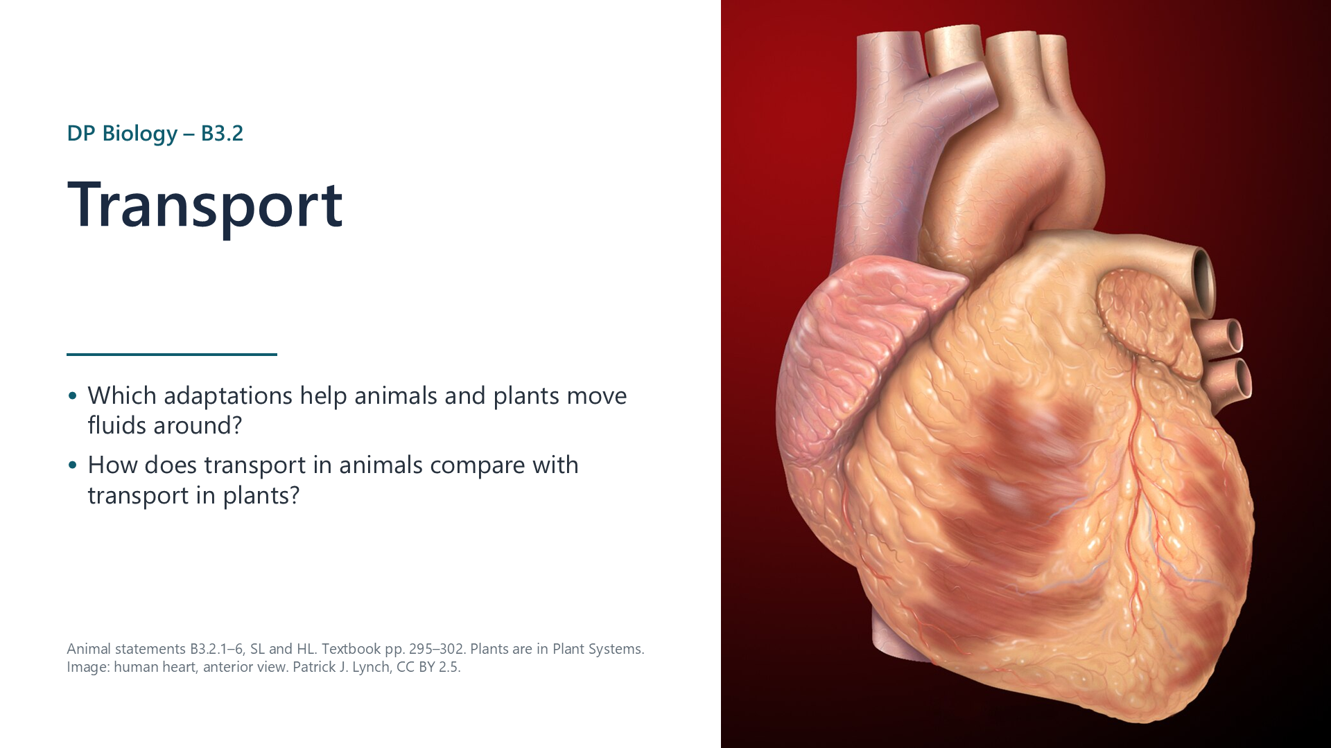
Slide 1 of 23

Notes

Written for this site from the course textbook and class slides. Use the page numbers to read the book's fuller account. Water transport in plants (B3.2.7–10) is in Plant Systems. Image credits are on slide 23.

Exchange

B3.2.1

How capillaries are adapted for exchange

p. 296

Capillaries are where materials pass between blood and tissue fluid. Each feature helps:

  • Branching gives a huge surface area, and no cell is far from a capillary.
  • Narrow diameter, about the width of one red blood cell, puts those cells right next to the wall.
  • Thin wall, a single layer of endothelial cells, makes the diffusion distance short.
  • Fenestrations, or small pores, occur in some capillaries where exchange has to be especially rapid, such as in the kidney and small intestine. Some plasma escapes to form tissue fluid.

Low pressure and slow flow also give more time for exchange.
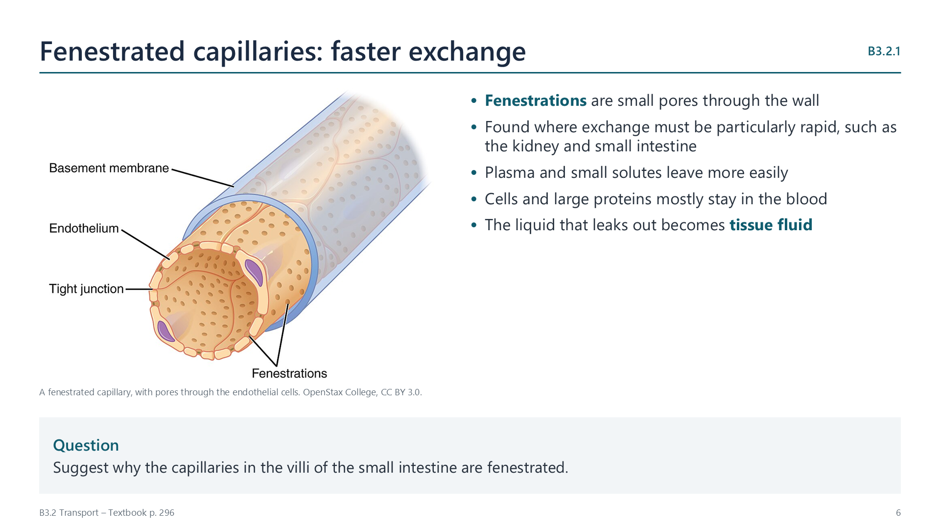
Slide: diagram of a fenestrated capillary beside key points

Arteries and veins

B3.2.2

The structure of arteries and of veins

pp. 296–297

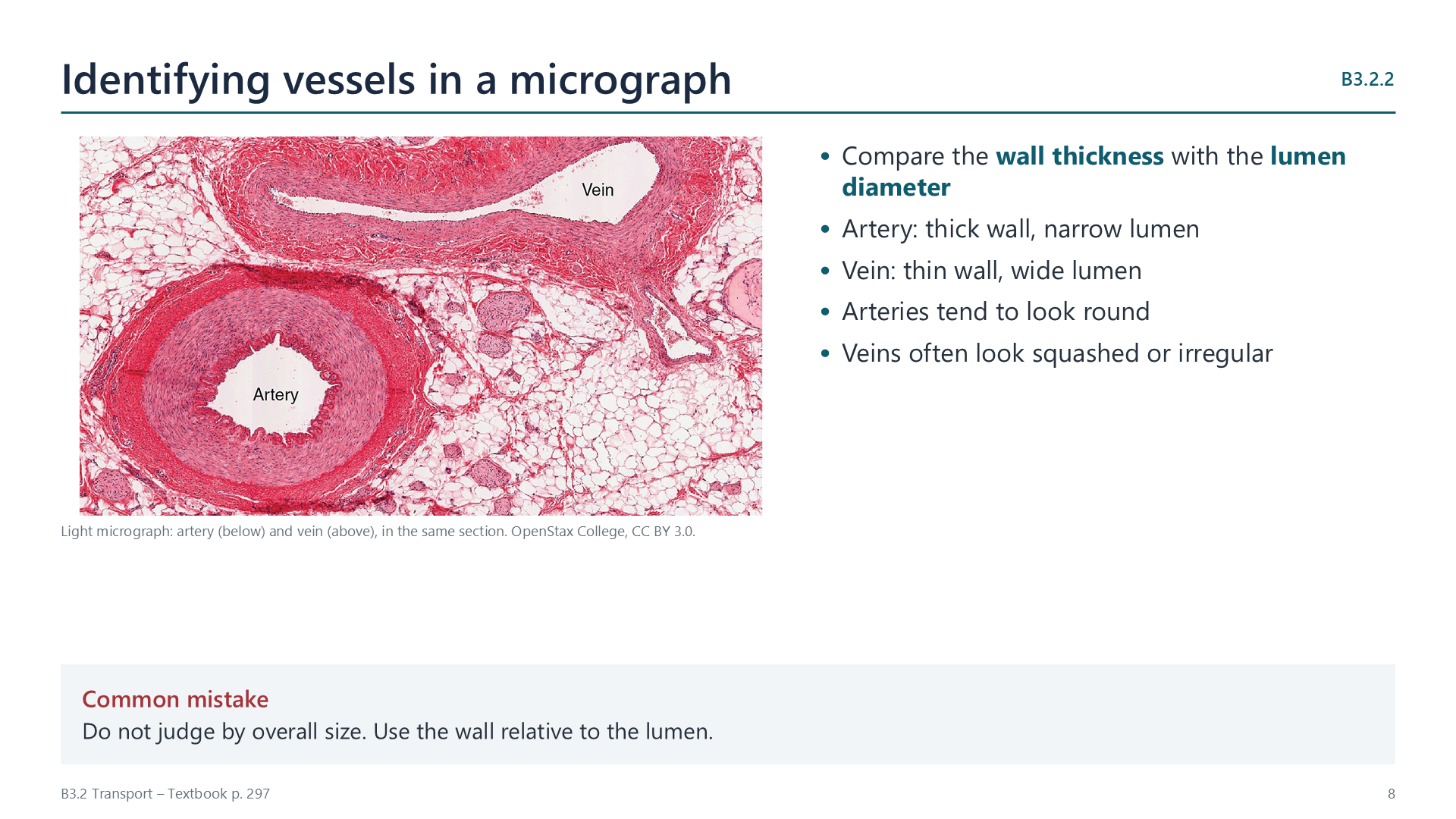
Both have three layers around a hollow lumen: the tunica intima (smooth endothelium), the tunica media (smooth muscle and elastic fibres) and the tunica externa (connective tissue with collagen).

Slide: light micrograph of an artery and a vein in the same section, with tips for telling them apart

In a micrograph, compare the wall with the lumen. An artery has a thick wall and a narrow lumen and tends to look round. A vein has a thin wall and a wide lumen and often looks squashed.

Watch out: size alone does not tell you which is which. Use wall thickness relative to lumen diameter.

B3.2.3

How arteries are adapted to carry blood away from the heart

pp. 297–298

Each heartbeat sends a surge of blood into the arteries at high pressure, reaching about 120 mm Hg in the aorta. The wall has two jobs:

  • Withstand the pressure: a thick wall of muscle, with collagen, prevents the artery from rupturing.
  • Maintain the pressure: elastic fibres stretch as the surge arrives, storing energy, and then recoil between beats, pushing the blood on. Contracting smooth muscle also narrows the lumen.

Further from the heart, arteries have less elastic tissue and more smooth muscle. Arterioles use this muscle to control how much blood reaches each capillary bed.

B3.2.4

Measuring pulse rate

pp. 298–299

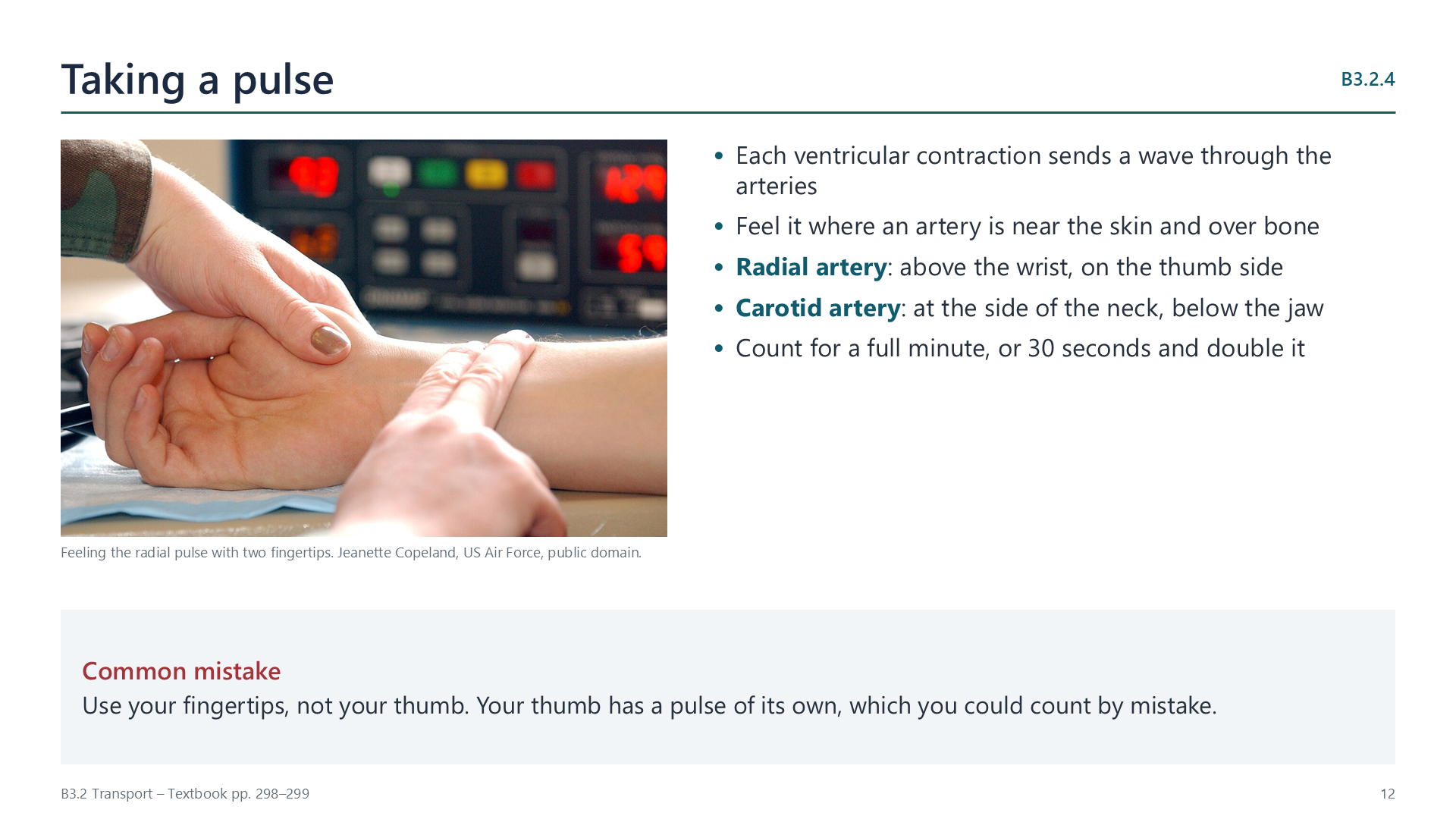
Each wave of blood from the ventricles stretches the arteries, and you can feel this where an artery runs near the skin and over bone: the radial artery above the wrist, or the carotid artery in the neck.

Press gently with two fingertips. Count for a full minute, or count for 30 seconds and double it. Do not use your thumb, as it has its own pulse.

Digital devices such as fitness watches shine an LED through the skin and detect changes in blood flow. They record continuously but can vary in accuracy.

Slide: photograph of two fingertips on a wrist beside instructions for taking a pulse
B3.2.5

How veins are adapted to return blood to the heart

p. 299

By the time blood reaches a vein it has lost nearly all its pressure, at about 5 mm Hg, and has no pulse. Backflow is a real risk, so veins rely on two features:

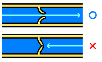
  • Valves: blood moving to the heart pushes the flaps aside. If it flows backwards, it fills the pockets and the flaps close.
  • A thin, flexible wall: contracting skeletal muscles can squeeze the vein, pushing blood along it.

The wide lumen also reduces friction. This is why walking helps blood to return from the legs.

When arteries become blocked

B3.2.6

What blocks coronary arteries and what follows

pp. 300–302

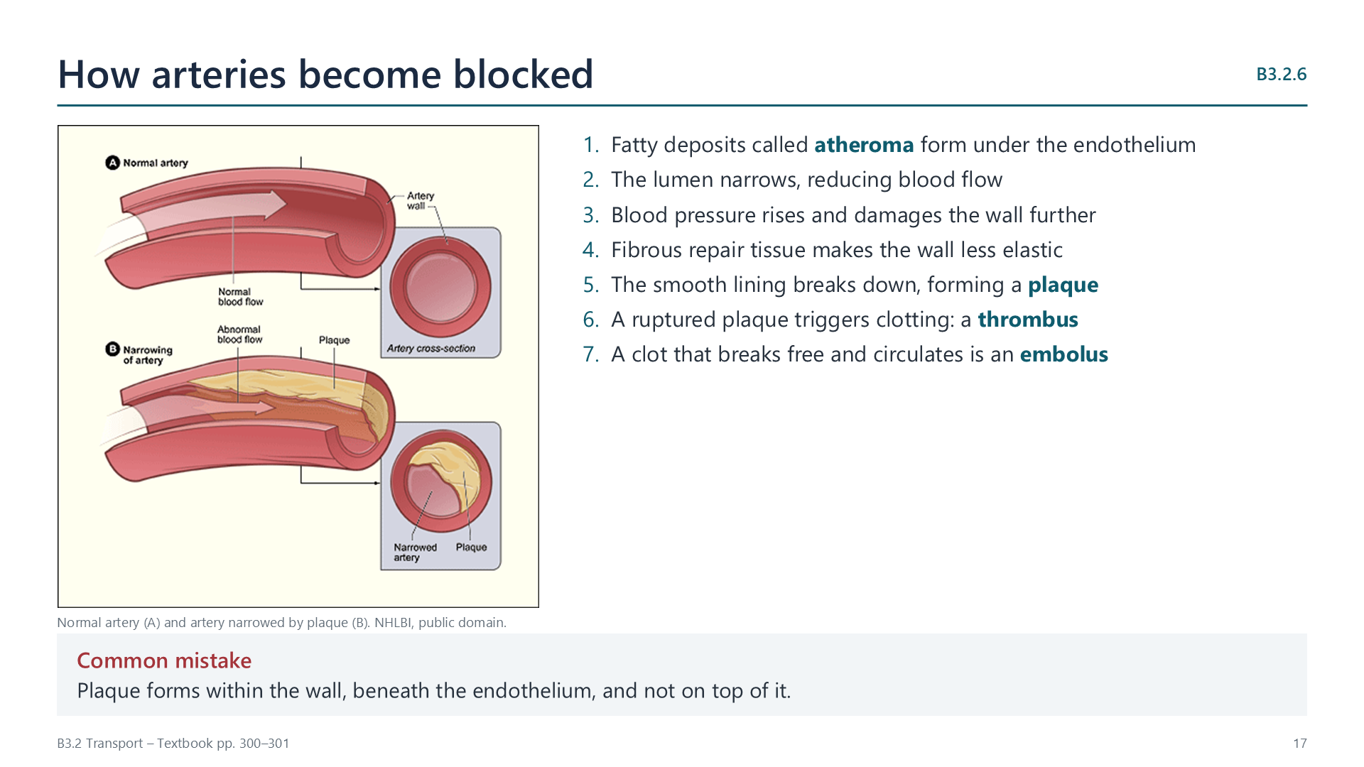
Coronary arteries branch from the aorta and supply heart muscle. Occlusion means that a vessel is blocked or closed.

  1. Fatty deposits, or atheroma, form under the endothelium.
  2. The lumen narrows and blood pressure rises, which damages the wall further.
  3. Fibrous repair tissue makes the wall less elastic.
  4. The lining breaks down, forming a plaque.
  5. A ruptured plaque triggers clotting, and the clot is a thrombus. If it breaks free it is an embolus.
Slide: a normal artery compared with one narrowed by plaque, beside the stages of atherosclerosis

Consequences: a narrowed artery can cause chest pain, or angina, during exercise. A full block deprives muscle downstream of oxygen, so it dies. If enough muscle is lost, the heart cannot pump effectively. This is a heart attack, or myocardial infarction.

Evaluating data: studies across populations link coronary heart disease with factors such as saturated fat, smoking and high blood pressure. When you evaluate this evidence, look at sample size, spread of data, whether other variables were controlled, and how the data were collected. A strong correlation does not prove a cause.

Watch out: plaque forms within the artery wall, beneath the endothelium. It does not simply stick on top.

Videos

Select a video to play it here. Select it again to close it.

Note-taking sheet

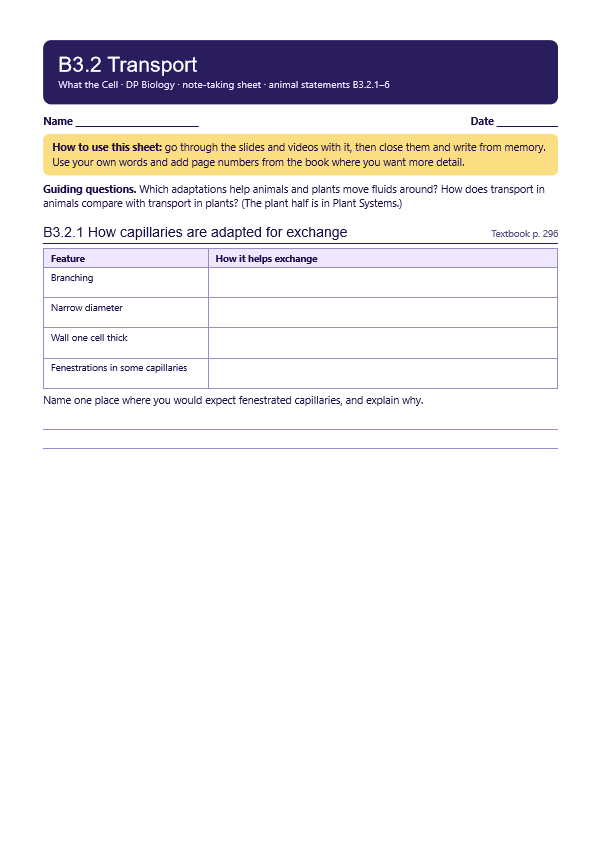
Print this guide and complete it as you work through the slides and videos. It asks for your own explanations, so close the notes while you try each section.

First page of the note-taking sheet

Quick self-check

The self-check needs JavaScript to run.

Practice questions

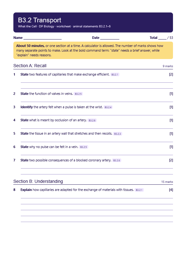
A printable worksheet covering all six statements, from quick recall to data questions and longer answers. It has 53 marks and takes about 50 minutes, or you can do one section at a time.

Your teacher has the mark scheme.

First page of the worksheet

Data-based questions

Questions built on realistic experimental data, in the style of Paper 1B and Paper 2. Work them on paper and show your working. 7 marks in total. Your teacher has the mark scheme.

Returning blood from the legs

7 marks
Valve in a vein
Photo: made by Was a bee., Public domain, Wikimedia Commons.

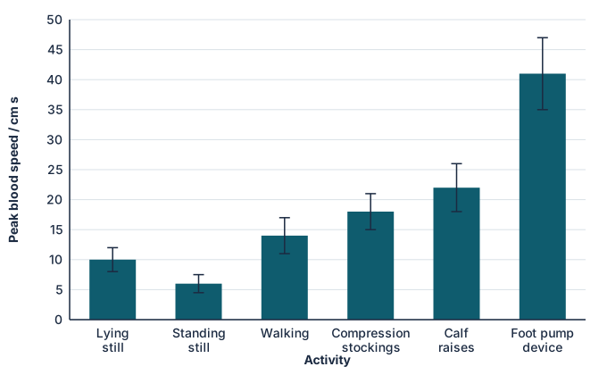
Blood in leg veins returns to the heart partly because contracting leg muscles squeeze the veins, and valves stop it flowing back. The peak speed of blood in a vein behind the knee was measured in 24 adults during six activities. Lying still was the control. Error bars show ± 1 SD.

Bar chart of peak venous blood speed. Lying still 10, standing still 6, walking 14, compression stockings 18, calf raises 22, foot pump device 41 centimetres per second.
  1. Identify the activity that reduces blood speed compared with the control. [1]
  2. Suggest a reason for this reduction. [1]
  3. Calculate the percentage increase in blood speed with compression stockings compared with lying still. [1]
  4. Determine the difference in blood speed between lying still and using the foot pump device. [1]
  5. People on long flights are advised to move their legs regularly. Discuss the benefits of this, using the data. [3]

Practical

Pulse rate and exercise

Lab30 minB3.2.4

You will need: Stopwatch, step or chair, results table

  1. Measure resting pulse at the wrist for 30 seconds and double it
  2. Do two minutes of gentle stepping
  3. Measure pulse immediately, then every minute until it returns to resting level
  4. Plot pulse rate against time and describe the recovery

Safety: Volunteers only. Anyone with a heart or breathing condition should act as timer. Stop if unwell.

Carry this out with your teacher's supervision and follow your school's lab rules.

Nature of science and TOK

Syllabus statements

Summarised in our own words. Use each code to find the exact wording in your textbook or the IB subject guide.

  • B3.2.1How capillaries are adapted to exchange materials with blood's surroundingsp. 296
  • B3.2.2The structure of arteries and of veinsp. 296
  • B3.2.3How arteries are adapted to carry blood away from the heartp. 297
  • B3.2.4Measuring pulse ratep. 298
  • B3.2.5How veins are adapted to return blood to the heartp. 299
  • B3.2.6What blocks coronary arteries and what followsp. 300

HL extension

More of B3.2 in this course

© 2026 Akash Parthi Bendran. Please ask before using or sharing any resources from this site: see permission to use.