What the Cell
B3.2

Transport

Plant
2 hours4 statementsTextbook pp. 303–311

Guiding questions

  1. Which adaptations help animals and plants move fluids around?
  2. How does transport in animals compare with transport in plants?

Slides

Slide 1 of 14

Notes

Written for this site from the course textbook and class slides. Use the page numbers to read the book's fuller account. Transport in animals (B3.2.1–6) is in Body Systems. Image credits are on slide 14.

Moving water

B3.2.7

How transpiration moves water from roots to leaves

pp. 303–307
  1. Water evaporates from cell walls in the leaf.
  2. More water is drawn out of xylem vessels and through cell walls by capillary action.
  3. This generates tension, or negative pressure, in the xylem.
  4. Cohesion between water molecules keeps the column continuous, so the tension pulls water up from the roots.

Adhesion between water and hydrophilic cell walls helps too. The plant spends no ATP on this movement, because the Sun supplies the energy for evaporation.
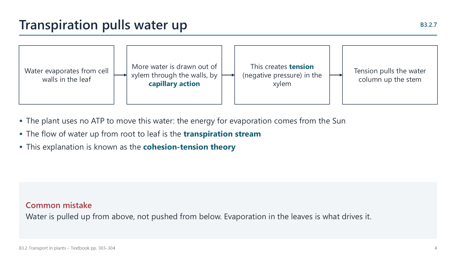
Slide: four steps from evaporation in the leaf to water being pulled up the stem

Evidence for tension: trunks of trees shrink slightly in the day, when transpiration is fastest. If a xylem vessel is pierced, air is drawn in and water does not spurt out.

Watch out: water is pulled from above, not pushed from below.
B3.2.8

How xylem vessels are adapted to carry water

p. 308
FeatureHow it helps
No cell contentsUnimpeded flow
Incomplete or absent end wallsContinuous tubes, with unimpeded flow
Walls strengthened with ligninWithstand tension without collapsing
PitsAllow water to enter and leave

Where the tissues are

B3.2.9

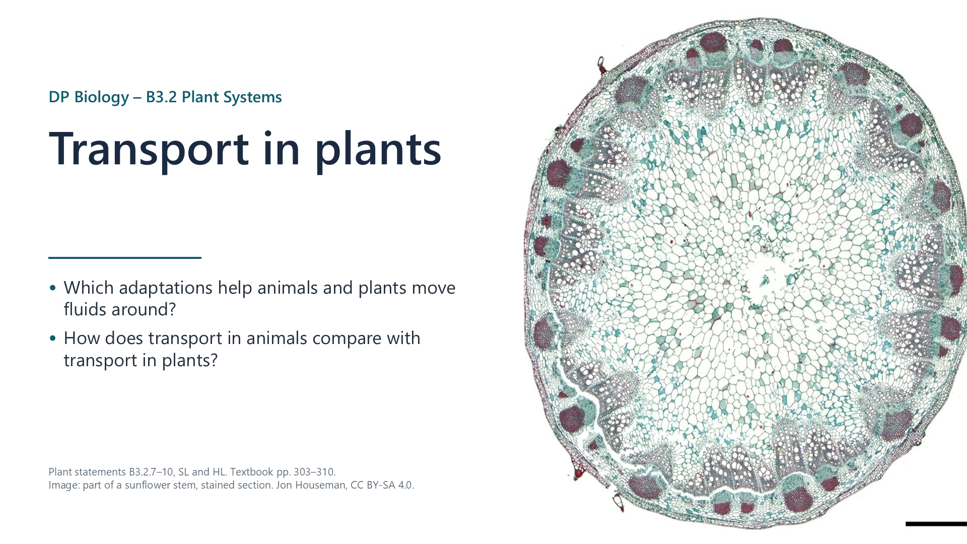
How tissues are arranged in a dicot stem

pp. 308–309

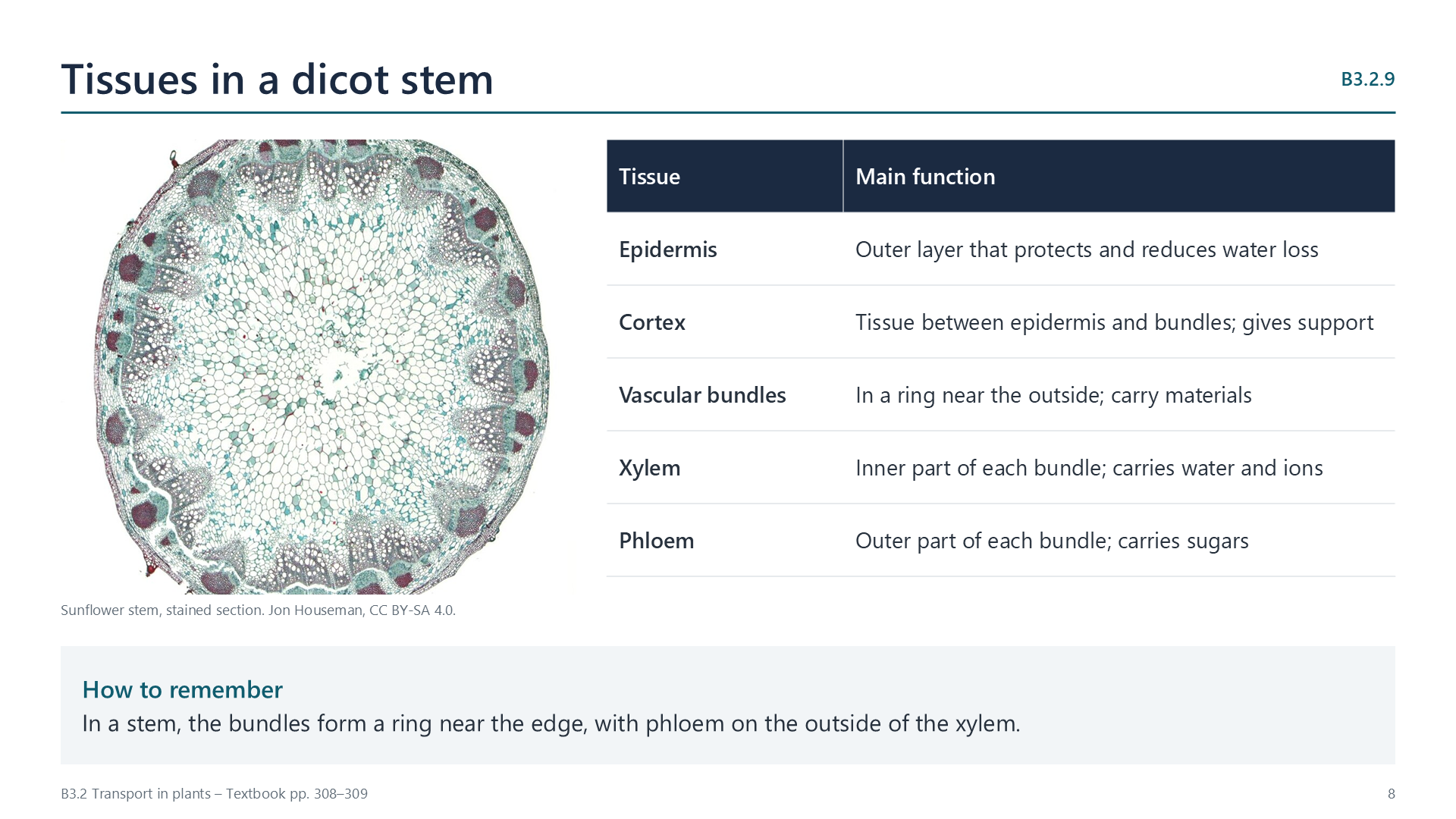
The epidermis forms the outer layer, and the cortex lies inside it. Vascular bundles form a ring near the outside. In each bundle, xylem carries water and ions towards the centre of the stem and phloem, which carries sugars, is on the outer side.

Slide: micrograph of a sunflower stem beside a table of tissues and functions
B3.2.10

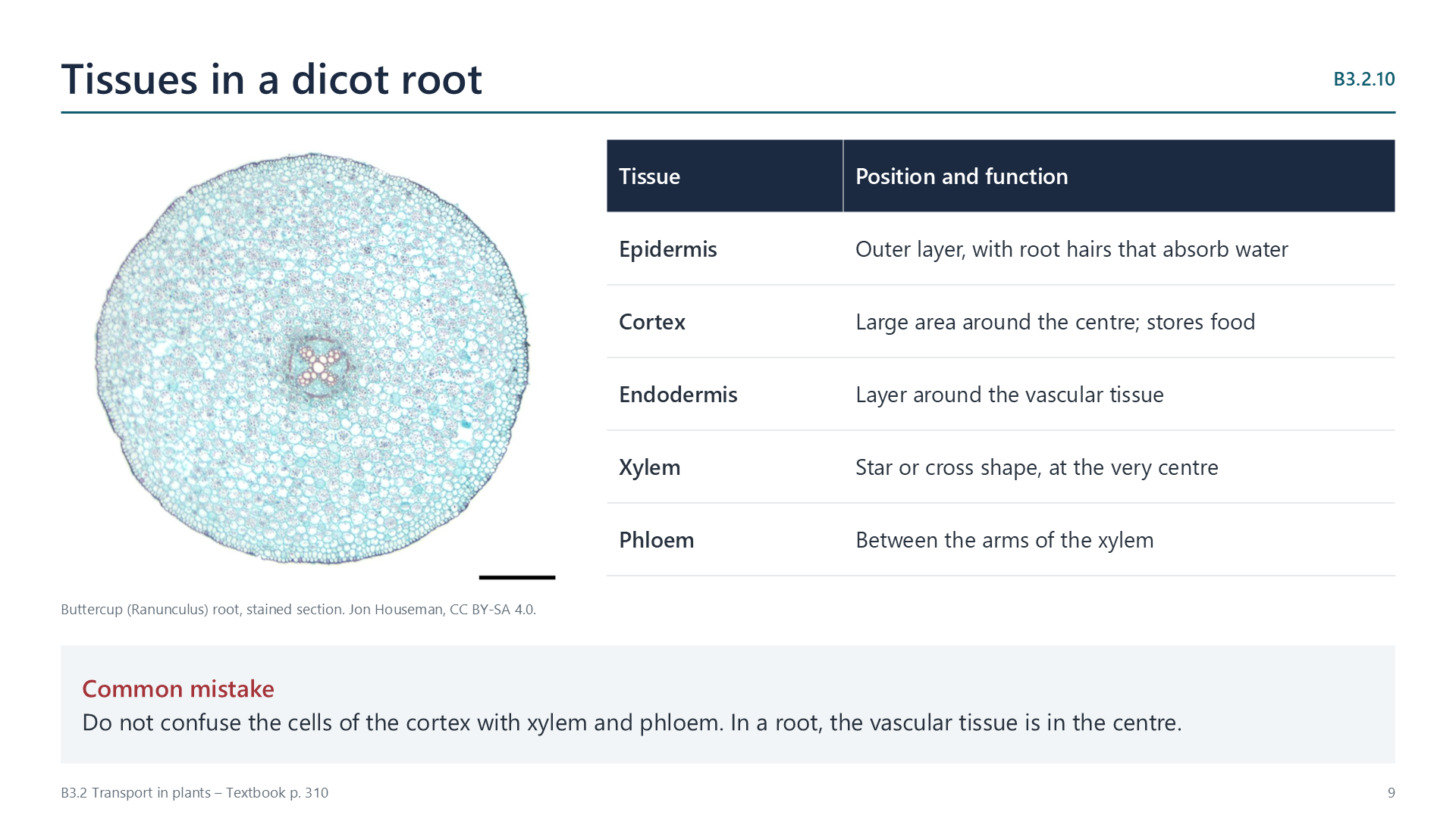
How tissues are arranged in a dicot root

p. 310

The epidermis has root hairs for absorbing water. The large cortex surrounds the central vascular tissue. Xylem forms a star or cross shape at the very centre, and phloem lies between its arms.

When you draw either plan diagram, show the outlines of tissues, not cells. Keep to the proportions of the micrograph, and label each boundary.

Slide: micrograph of a buttercup root beside a table of tissues and functions

Watch out: do not confuse cortex cells with xylem and phloem. In a root, the vascular tissue is in the centre.

Videos

Select a video to play it here. Select it again to close it.

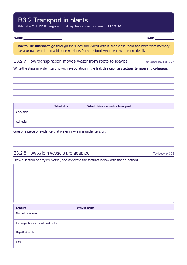
Note-taking sheet

Print this guide and complete it as you work through the slides and videos. It asks for your own explanations, so close the notes while you try each section.

First page of the note-taking sheet

Quick self-check

The self-check needs JavaScript to run.

Practice questions

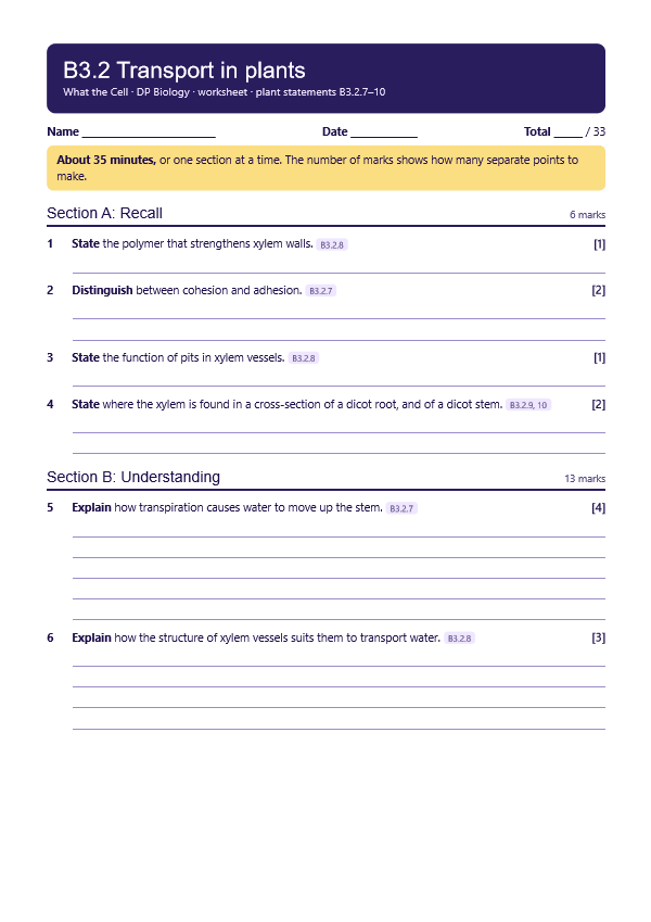
A printable worksheet covering all four statements, from quick recall to data questions. It has 33 marks and takes about 35 minutes, or you can do one section at a time.

Your teacher has the mark scheme.

First page of the worksheet

Practical

Tracing water through xylem

Lab30 min, plus a few hoursB3.2.7, B3.2.8, B3.2.9

You will need: Celery stalk with leaves, food dye, beaker, scalpel, hand lens or microscope

  1. Stand a freshly cut stalk in dyed water for a few hours
  2. Cut thin cross-sections from different heights
  3. Observe where the dye appears
  4. Draw the section and label the xylem, then explain how the water rose

Safety: Cut away from your fingers on a board. Food dye stains clothes.

Carry this out with your teacher's supervision and follow your school's lab rules.

Nature of science and TOK

Syllabus statements

Summarised in our own words. Use each code to find the exact wording in your textbook or the IB subject guide.

  • B3.2.7How transpiration moves water from roots to leavesp. 303
  • B3.2.8How xylem vessels are adapted to carry waterp. 308
  • B3.2.9How tissues are arranged in a cross-section of a dicot stemp. 308
  • B3.2.10How tissues are arranged in a cross-section of a dicot rootp. 310

HL extension

More of B3.2 in this course

© 2026 Akash Parthi Bendran. Please ask before using or sharing any resources from this site: see permission to use.